Dr Sandeep Kumar’s Clinic offers best surgery services for Abdominal pain in Lucknow.
- A clinical syndrome of severe abdominal pain
- Onset variable from minutes to hours to weeks
- Variable severity – mild to moderate to unbearable severe
- Acute first time / Acute exacerbation of a chronic pain / Acute intermittent / Acute recurrent
- All age groups and sex
- Accompaniments : loose motion / diarrhoea / tensmus; nausea / vomiting; haematemesis / melena / haematochezia; fever, toxic symptoms, radiations, haemodynamic instability, sepsis / shock, metabolic disorders,

Clear cut signs of peritonitis – superficial tenderness, rigidity and hyperesthesia
Developing signs of peritonitis
Completely and mostly haemodynamically stable
Haemodynamic in stability imminent / haemodynamically unstable – shock
Haemotological tests – normal / abnormal – causal / inferential / incidental
Biochemical tests – normal / abnormal – causal / inferential / incidental
USG findings – corroborative to history and examination
Incidental USG finding – unrelated to physical examination and history
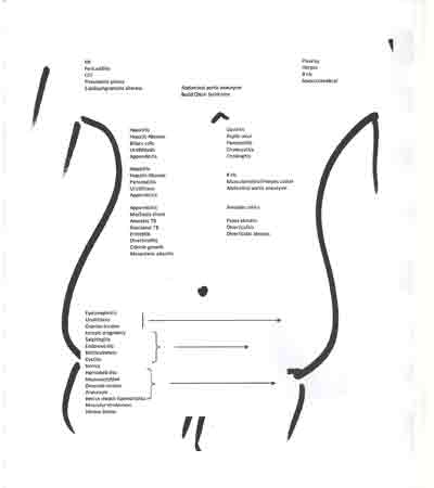
Therefore, acute abdomen revisited as 3 clinical entities :
Who are you? Scenario
- Family Physician who will treat medically only and haemodynamically stable acute abdomen
- Attending Physician who would treat medically but normal conditions how so ever serious
- Primary Surgeon ready to take up selected doable acute abdomen
- Attending Surgeon who has to deal with all cause acute abdomen
| Diagnosed | Undiagnosed | Pseudo-diagnosed |
| Face value diagnosis obvious Tests usually USG findings significantly corroborate with clinical impression Medical or surgical treatment Debates on treatment – immediate versus delayed operations on appendicitis, cholecystitis, pancreatitis, intra-abdominal abscesses etc |
Diagnosis comes easy on history, physical examination, blood tests, biochemistry, x-ray, ultrasound etc
Diagnosis remains enigmatous despite above tests |
Often wrongly diagnosed or treated because of an indexed abnormality in one of the tests specially ultrasound for example showing incidental gall stones, fibromyoma uterus, hamartoma liver, benign renal cyst etc and these findings do not corroborate with clinical symptoms and signs Patient does not get good relieve after treatment of incidentinoma |
Case Scenario 1 : Ms AP 26 yrs, bank probationary officer
PC : Acute abdomen – recurrent – 6 months, no fever, 4 kg weight loss. Sometimes vomiting
Physical Examination : Generally well, pallor, haemodynamically fully stable
No visceromegaly, some visible peristalsis and exaggerated bowel sounds
Investigations :
Haemotology – WNL
Biochemistry – WNL
X-rays – plain abdomen erect films – SAIO
USG – WNL
Triphasic triple contrast coronal section CT abdomen – dilated jejunal loops, suspicious intestinal stricture
Operative finding : Two tight circumferential jejuno-ileal area strictures 10 cms apart, histopath – chronic granuloma, segment of intestine ressected bowel plumbed end to end.
Follow up : Ten months ATT, 1 year full resolution
Case Scenario 2 : Ms PT 32 yrs married female house worker
PC : Post caesarian acute abdomen – this was her 3rd C-section
Recurrent acute abdomen – 1 month, fever, 2 kg weight loss, vomiting, well being not attained post C-section
Physical Examination : Generally well, pallor, haemodynamically fully stable
No visceromegaly, some visible peristalsis and exaggerated bowel sounds
Investigations :
Haemotology – WNL
Biochemistry – WNL
X-ray – fluid levels in erect film
USG – WNL
Triphasic triple contrast coronal section CT abdomen – dilated jejunal loops, suspicious intestinal stricture
Operative finding : Terminal ileum engaged in uterine stump. Huge amount of chromic catgut, products of conception, clotted blood, organized frozen pelvis.
Careful 2 hours dissection, bowel dis-engagement, small bowel walk DJ to ileo-caecal, revised closure vaginal stump and conservation of ovaries on both sides
Follow up : Six months – full recovery

